Our Services

For everyday care or life-changing care, you can count on us to keep you and your loved ones safe and healthy

Spine

The bones, muscles, tendons, and other tissues that reach from the base of the skull to the tailbone.

Read More

Arthroscopy

The bones, muscles, tendons, and other tissues that reach from the base of the skull to the tailbone.

Read More

Joint Replacement

The bones, muscles, tendons, and other tissues that reach from the base of the skull to the tailbone.

Read More

Infertility

The bones, muscles, tendons, and other tissues that reach from the base of the skull to the tailbone.

Read More

All Gynaecological Problems

The bones, muscles, tendons, and other tissues that reach from the base of the skull to the tailbone.

Read More

TLH

The bones, muscles, tendons, and other tissues that reach from the base of the skull to the tailbone.

Read More

What Is Avascular Necrosis?

Avascular necrosis (AVN) is the death of bone tissue because of a loss of blood supply. You may additionally hear it known as osteonecrosis, aseptic necrosis, or ischemic bone necrosis. If it isn’t treated, AVN will cause the bone to collapse. AVN most frequently affects your hip. Different common sites are the shoulder, knees, and ankles.

  • Symptoms:

In its early stages, AVN sometimes doesn’t have symptoms. As the sickness gets worse, it becomes painful. At first, it’d solely hurt once you place pressure on the affected bone. Then, the pain might become constant. If the bone and encompassing joint collapse, you will have severe pain that makes you unable to use your joint. The time between the primary symptoms and bone collapse will vary from many months to quite a year.

  • Causes And Risk Factors:

Things that will create avascular necrosis a lot of likely includes:

  • Alcohol:

Several drinks every day will cause fat deposits to make in your blood, which lowers the blood provided to your bones.

  • Bisphosphonates:

These medications that boost bone density may lead to osteonecrosis of the jaw. This might be a loft possibility if you’re taking them for multiple myeloma or Metastatic Breast Cancer.

  • Medical Treatments:

Radiotherapy for cancer can weaken bones. Different conditions connected to AVN embody organ transplants, like kidney transplants.

Steroid long. Doctors don’t understand why however the longtime use of medicines like prednisone will cause AVN. They assume the meds will raise fat levels in your blood, which lowers blood flow.

  • Trauma:

Breaking or dislocating a hip will harm near blood vessels and cut the blood provided to your bones. AVN might affect the 200th or a lot of individuals who dislocate a hip.

Blood clots, inflammation, and harm to your arteries. All of those will block blood flow to your bones.

Other Conditions Related To AVN:

  • Decompression illness, that causes gas bubbles in your blood
  • Diabetes
  • Gaucher sickness, within which a fatty substance collects within the organs
  • HIV
  • Long-term use of medication known as bisphosphonates to treat cancers like multiple myeloma or breast cancer, which might cause AVN of the jaw.
  • Pancreatitis, inflammation of the duct gland
  • Radiation therapy or chemotherapy
  • Autoimmune diseases like lupus
  • Sickle cell sickness

Who Gets Avascular Necrosis?

As many as 20,000 individuals develop AVN every year. Most are between ages twenty and fifty. For healthy individuals, the chance of AVN is little. Most cases are the results of AN underlying ill health or injury.

Avascular necrosis diagnosing

Your doctor can begin with a physical exam. They’ll proceed with your joints to ascertain tender spots. They’ll move your joints through a series of positions to ascertain your variety of motion. You may get one amongst these tests to know for what’s inflicting your pain:

  • Bone scan:

At first in your vein, the doctor injects radioactive materials. It travels to spots wherever bones are wounded or healing and shows the informed image.

  • MRI and CT scan:

These offer your doctor careful pictures showing early changes in the bone which may be a symptom of AVN.

  • X-rays:

They’ll be normal for the early stages of AVN however will show bone changes that seem afterward.

  • Treatment

Treatment for AVN is to boost the joint, stop bone harm, and ease the pain. The simplest treatment can rely on a variety of things, like:

  • Your age
  • Stage of the sickness
  • Location and quantity of bone harm
  • Cause of AVN

If you take avascular necrosis early, treatment might involve taking medications to relieve pain or limiting the use of the affected area. If your hip, knee, or ankle joint is affected, you will like crutches to require weight off the damaged joint. Your doctor might also suggest range-of-motion exercises to assist keep the joint mobile.

  • Medications:

If the doctor is aware of what’s inflicting your avascular necrosis, treatment can embody efforts to manage it. This will include:

  • Blood Thinners:

You’ll get these if your AVN is caused by blood clots.

  • Nonsteroidal Anti-inflammatory Drugs (NSAIDs):

These can facilitate pain.

  • Cholesterol Drugs:

They cut the quantity of cholesterol and fat in your blood, which might facilitate stopping the blockages that cause AVN.

  • Surgery

While these non-surgical treatments might block the avascular necrosis, the general public with the condition eventually would like surgery. Surgical choices include:

  • Bone Grafts:

Removing healthy bone from one part of the body and victimization it to interchange the broken bone

  • Osteotomy:

Cutting the bone and changing its alignment to alleviate stress on the bone or joint

  • Total Joint Replacement:

Replacing the damaged joint with an artificial joint

  • Core Decompression:

Removing a part of the inside of the bone to alleviate pressure and permit new blood vessels to make

  • Vascularized Bone Graft:

Using your own tissue to build diseased or damaged hip joints. The surgeon removes the bone with the poor blood, then replaces it with the blood-vessel-rich bone from another site, like the calf bone.

  • Electrical Stimulation:

An electrical current might jump-start new bone growth. Your doctor may use it throughout surgery or offer you a special gadget for it.

  • Caring For AVN At Home

You can do this stuff to help:

Rest.

  • Stay off the joint:

This will facilitate slow harm. You may have to be compelled to wait on physical activity or use crutches for many months.

  • Exercise:

A physiotherapist can show you the correct moves to induce a variety of motions back in your joint.

  • Prevention

To lower your risk of AVN:

  • Cut back on alcohol.
  • Heavy drinking may be a leading risk issue for AVN.
  • Keep your cholesterol in restraint.
  • Little bits of fat are the foremost common factor that blocks blood provided to your bones.
  • Use steroids carefully.
  • Your doctor ought to keep tabs on you whereas you’re taking these medications. Allow them to understand if you’ve used them in the past. Taking them over and yet again will worsen bone harm.
  • Don’t smoke.
  • It boosts your AVN risk.

The prognosis for avascular necrosis:

More than half the people with this condition would like surgery within three years of diagnosing. If a bone collapses in one of your joints, you’re out of possibilities to have it happen in another.

How Balloon Kyphoplasty Works – A Detailed Description of Balloon Kyphoplasty, Side Effects, And its Risks.

balloon kyphoplasty

Balloon kyphoplasty is the same as vertebroplasty. It is a minimally invasive procedure treatment for spinal compression fractures (also known as vertebral compression fractures of VCFs). The main purpose of this treatment is to relieve pain, restore vertebral height, and correct kyphosis. Balloon Kyphoplasty is the modified technique of vertebroplasty; the only main difference is that balloon kyphoplasty uses as the suggest a small balloon that gently lifts and put the bone fragments into their correct position. Balloon kyphoplasty can be effective in treating people whose vertebrae collapse due to osteoporosis,(mainly a disease that causes loss of bone density) or whose bones are weakened by cancer. This treatment is used to recover recent fractures. Kyphoplasty increases the patient’s functional abilities, allow gain strength and to perform the previous level of activity, and prevent further vertebral collapse. The procedure is usually successful at alleviating the pain caused by a compression fracture; many patients feel significant relief almost immediately

There is no upper age limit for this treatment. And the best part of balloon kyphoplasty is that there are more benefits than its side-effects.

The procedure of balloon kyphoplasty:

Your surgeon performs these steps for balloon kyphoplasty:

  • Firstly, you maybe given some general anesthesia and antibiotic to prevent infection.
  • Then the surgeon inserts a hollow needle (trocar) into your skin. With the help of fluoroscopy, (a type of X-ray), they guide the needle through your muscles and into the correct position in your bone.
  • The next step is to insert an inflatable balloon into the trocar.
  • Once it is in the position the balloon is then inflated to create the space required for the bone cement.
  • After space has opened up, the mixture is injected to fill it up. Imaging tests will help the surgeon confirm that the mixture is distributed properly.
  • Once the cement is in place, gently the needle is removed.
  • The area is bandaged. Stitches won’t be necessary.
  • A CT-scan may be used at the end of the procedure to check and verify the distribution of the cement.

After the treatment, a patient must be advised to stay in a recovery room for a short time usually a day. Immediately after the procedure, you may have some soreness or pain which can be relieved after 48hrs of your treatment. It is not a time-consuming procedure it usually takes half-per level.

Balloon kyphoplasty risks:

As we know every procedure has some risks and benefits.

  • If we talk about balloon kyphoplasty this treatment is not advisable for back arthritis, herniated disks, or curvature of the spine surgeon in Delhi due to scoliosis. It might have some serious consequences. However, the risk or complication rate for Balloon Kyphoplasty is very low, as with most surgical procedures serious adverse events, some of which can be fatal, can occur, including heart attack, cardiac arrest (heart stops beating), stroke, and embolism (blood, fat or cement that migrates to the lungs or heart).
  • Some other risks include leakage of bone cement into the muscle and tissue surrounding the spinal cord and nerve injury that can, infection, in some rare instances, it causes paralysis; leakage of bone cement into the blood vessels resulting in damage to the blood vessels, lungs and/or heart.
  • increase the pain- sometimes balloon kyphoplasty worsens the pain. It either fails to relieve the pain or results in worse pain or other symptoms. For example, if bone cement leaks onto a nerve root or the spinal cord, it could potentially cause worsened symptoms of pain, tingling, numbness, and/or weakness. This happens in some very rare case of around 5% of the patients undergoing balloon kyphoplasty does not get relief from the pain.

It is very important to consult the Best Orthopedic Doctor in Delhi NCR before going through the treatment as balloon kyphoplasty is not for everyone.

Balloon kyphoplasty Side-effects:

  • redness of the place where the treatment has been done.
  • soreness
  • anesthetic risks,
  • surgical mistakes,
  • There is a possibility of infection
  • It can cause bleeding where the needle is penetrated
  • In some cases the procedure causes another fracture in the spine or ribs.
  • nerve damage from the procedure can lead to numbness, weakness, or tingling
  • pulmonary embolism.

Hence, if you wish to go for this treatment this is all you need to know. It is highly recommended to consult the doctor before undergoing balloon kyphoplasty to prevent serious consequences.

Plantar Fasciitis Treatment

If you are feeling the pain at the bottom of your heel, it might be due to Plantar Fasciitis. Plantar Fascia is a weblike, thick ligament that connects the front to the heel of your foot. It helps you in walking and also acts as a shock absorber. It also supports the arch of your foot while you walk. Dr. Gaurav Khera is a Best Joint Replacement Surgeon offering world-class surgeries.

What is the Planter fasciitis:

One of the most common complaints of Orthopaedic Surgeon is that of Plantar Fasciitis. In our day to day life, our Plantar Fascia faces a lot of wear and tear, and high pressure on it can even damage your ligaments. It generally causes inflammation in heels and also leads to stiffness.  The cause of discomfort in Plantar Fascia is still not known.

What cause planter fasciitis

The causes that are responsible for the Plantar Fasciitis are stated below.

  • The highest risk of Plantar Fasciitis is to the active men and women between the ages group of 40 to 70 years.
  • The problem is quite common in women when compared to men.
  • The women in late pregnancy might experience the pain of Plantar Fasciitis.
  • Those who are overweight have higher chances of suffering from Plantar Fasciitis. The main reason behind it is that higher pressure is experienced due to the gain of weight, it might lead to the wear and tear of ligaments.
  • Those who are long-distance runners can also develop this problem.
  • Those who have active work of standing on feet might also experience this problem.
  • Other than these problems, the person with structural foot problems might develop this problem.
  • The person wearing shoes with soft soles and poor arch experience the pain of Plantar Fasciitis.

Generally, people believe that the Plantar Fasciitis cause is the result of heel spurs, but this is not the actual cause of the problem.

Symptoms of Plantar Fasciitis

Plantar Fasciitis is the problem where the person suffers from pain in the bottom of the heel or might experience pain at the midfoot area. Generally, the pain is experienced in one foot, but in some cases, the pain may be experienced in both the feet.

The symptoms of this problem include pain.

  • The pain develops with time and is not an immediate one, and can be mild or intense.
  • The people might experience a burning sensation and the pain that extends outward from the heel.
  • The person suffering from Plantar Fasciitis will experience severe pain early in the morning, just after the person keeps his/her step out of the bed.
  •  The person suffering from Plantar Fasciitis will experience difficulty in climbing the steps due to stiffness in the heels.
  •  The person doesn’t experience the pain while doing the activity, instead, the pain is felt as soon as they stop working.

Testing and Diagnosis of Plantar Fasciitis

When it comes to testing and doing the diagnosis of the problem, the doctor might opt for different ways to diagnose the problem.

  • In order to know the exact location of the pain, the doctor might perform a physical examination, that will help him in examining the tenderness in the foot.
  • The doctor can also put pressure on the foot, in order to observe, if the pain worsens or not.
  • He might note if the person is experiencing redness or swelling.
  • The doctor will also examine the strength of muscles and the health of the nerves, by checking reflexes, muscle tone, sense of touch and sight, coordination, and balance.
  • The doctor can also suggest an X-ray or an MRI scan to know the reason behind the pain is Plantar Fasciitis and not any fracture.

How to cure planter fasciitis in one week

The treatment of Plantar Fasciitis includes both home remedies and getting it treated by the doctor.  The person suffering from this problem can use home remedies like icing, rest, using braces, and anti-inflammatory drugs. This is the first and foremost step to treat the problem. In case the pain is still very severe than it is recommended to consult the doctor to get the feet treated, as there are chances of ligament damage, for which the person needs to take an injection.

The doctor in order to treat your pain might use an ultrasound device so that the correct place to inject the injection could be identified. He can also apply corticosteroids to apply an electric current, that will allow the sterile to pass into muscle in the arch of the foot or heel.

The most effective way to reduce the pain of Plantar Fasciitis is to go for physical therapy. The therapist will tell the exercise to strengthen the muscle of the lower leg, which will help in balancing while walking.

In case the pain of the person is not getting relief from all these things, then the doctor might suggest extracorporeal shock wave therapy, which involves the bombarding by the sound waves to push the healing ligament.

The side effect of the treatment will include swelling, pain, numbness, and bruises

How to fix planter fasciitis

In case the patient is suffering from pain even after various ways, then another option to treat Plantar Fasciitis is surgery. This is done only when the case is severe and lasts for more than 6 to 12 months.

In this process, the surgeon partially removes the plantar fascia from the bone of the heel. It will definitely reduce the pain but will weaken the arch of the foot and the complete functioning might stop.

Surgery should be considered as the last option for treating your heel if other options fail to work. This is because it may lead to severe pain and can cause damage to the nerve.

How long does planter fasciitis last

The person suffering from this problem can also try the exercise for getting rid of the problem.

  • One can practice stretches that can provide relief from the pain. Because it helps in losing the muscle and providing relief to the pain in the heel.
  • The person suffering from Plantar Fasciitis should take a break from exercises like running and must opt for swimming and other activities, that will give time to get it to relax and hence will reduce the heel pain.
  • It is suggested to do stretches in order to make your heel relax and lower the pain.

How to get rid of planter fasciitis – Home Remedies for Plantar Fasciitis

 In order to reduce the pain and inflammation in the heel treating the Plantar Fascia ligament. The home remedies for treating it includes as listed.

  • The person can reduce the pain by applying the ice pack for around 10-15 minutes, for around three to four times a day. This will lead to a reduction in the swelling.
  • The person can also change the exercise or activity, that they do regularly and are responsible for the heel pain.
  • One can also go for the stretching exercise that relieves the pain of the heel.

Oils for Plantar Fasciitis

If the person wants to recover from the problem without going to the doctor, he/she might opt for the oils to reduce the pain of Plantar Fasciitis. The essential oils are a good option to get relief from the pain and inflammation caused by Plantar Fasciitis. These essential oils include-

  • lavender essential oil
  • rose essential oil
  • eucalyptus oil
  • lemongrass essential oil

It is suggested to use these oils by mixing it with coconut oil so that it becomes diluted. One can also inhale the essential oils through steam. Though the oils are not much beneficial in providing relief from the inflammatory caused by Plantar Fasciitis, if used properly might work for some, as there is no harm in using it to look for the results.

How to heal planter fasciitis quickly

If you want to avoid the problem of Plantar Fasciitis, then you can adopt a few things that will change your lifestyle. This change in lifestyle will help you in avoiding the problem to a larger extent.

  • It is suggested to wear good shoes that can provide good support to the arch of your foot.
  • It is also suggested to replace the shoes in a regular interval of time.
  • In case you are a runner, then you use to replace your shoes once you have run 400 to 500 miles. This should be the limit of a pair of shoes you use.
  • Try to go for exercise that does not always involve your foot to remain active.
  • Work hard to maintain your weight, as an increase in weight will surely lead you to face the problem of Plantar Fasciitis. Because if you are overweight it might lead to wear and tear of the ligaments.

Recovering from the Plantar Fasciitis

Generally, people recover from the problem within a few months of home treatment, which includes stretching, resting, and icing. It is also suggested to make use of tape to stabilize your foot, as it will limit the movement of the ligament. For this, you can opt for zinc oxide tape or kinesiology tape. This might prove effective for treating the foot temporarily.

The majority of the people get relief from home remedies and don’t have to opt for any kind of surgery. Though treating it might range from a few months to 2 years.

Arthroscopy – Knee and Shoulder Scopes

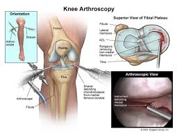
Arthroscopy is the ray of light for those who have painful and debilitating joint conditions. Joint arthroscopy, also known as knee and shoulder problem, is reckoned as one of the most effective and credible options for diagnosing and curing several orthopedic issues. It is a sophisticated method and your doctor may recommend you if you have inflammation or damaged a joint over time. Usually, it is done on the knee, shoulder, hip, wrist, ankle or elbow.

What is Arthroscopy?

Arthroscopy is the procedure that allows the orthopedic surgeon to look at the residing problem in your joint. The surgeon inserts a thin instrument into the joint through a small incision. It consists of a light and a camera that provides a magnified view of the joint on the computer screen. Hence the surgeon can effectively see the damage or deterioration and on the based on that can make an accurate diagnosis.

The advantage of arthroscopy

  • It is minimally invasive procedure
  • It provides faster recovery
  • It is less painful than open surgery
  • It has less or fewer complications

When to see an orthopedic doctor for arthroscopy

If you are grappled with the below-mentioned condition, you should consult to an orthopedic doctor and see if arthroscopy is an option for you:

  •  Joint inflammation
  • Pinched nerves
  • Shoulder instability (Condition in which cartilage that lines the rim of the shoulder tears, consequently shoulder joints become loose and may dislocate)
  • Rotator cuff problem in the shoulder
  • Arthritis or osteoarthritis
  • Misaligned kneecap
  • Baker’s cyst

How the arthroscopy procedure is performed?

Arthroscopy is recommended and beneficial for those suffering from rotator cuff injuries that are quite common these days and debilitating as well. Rotator cuff includes muscles and tendons surrounding the ball and socket joint of your shoulder. It is a minimally invasive procedure that offers reduced blood loss, fewer complication and faster recovery as compared to open surgery. Though rotator cuff injuries are considered to be difficult to treat tradition, however, with the advent of arthroscopy procedure, the surgeons are able to successfully treat or repair such issues.

During the procedure, the patient will be administered local or regional or at times general anesthesia as well so that the patient feels comfortable and does not feel pain. The surgeon will also inject saline into the joint in order to inflate the surgical area, to get explicit views and control bleeding.  

The surgeon makes  a small incision to insert the thin instrument  (arthroscope). He also makes several other small incisions to insert surgical tools. The light and camera attached with the instrument displays image on the screen that helps to guide the surgery. The procedure helps to inspect, diagnose and evaluate the problem in your joint. It also helps to perform arthroscopy surgery to treat or fix the problem.

As I mentioned that it is a less invasive procedure, however, if you knee or shoulder damage is quite prominent and the surgeon may conduct an open surgery to cure it.

Risks and Complications

Arthroscopy procedures are usually quite safe, however, like any other surgical procedure, it may have some complications and risks such as:

  • Impairment to the cartilage or ligaments
  • Blood clot in the leg
  • Allergic reaction to the anesthesia
  • Excessive bleeding
  • Joint stiffness
  • Injury to blood vessel or nerve

What happens after the arthroscopy?

The surgery is performed as an outpatient procedure. It means the patient can go home the same day. The incisions that are made during the procedure, may take several days or a week to heal. You may also experience some pain or discomfort for a weak or so. The consulting doctor may provide you the recovery and rehabilitation program and instruction to stabilize the joint and regain the strength and mobility after the surgery.

Action step

When you witness that your shoulder and knee condition is not responding to non-invasive treatment or methods including medication, physical therapy, consult your doctor that arthroscopy is ideal for you. Please contact us to schedule the appointment.

Knee Arthritis and High Tibial Osteotomy

Are you suffering from knee pain? Well, this pain can be caused by an injury or it may be due to wear and tear or by overuse. If the knee pain is a result of overuse, then it is advised that you take ample rest. This pain will subside as soon as you take rest. But, at times you may face persistent pain and this indicates that there is underlying medical condition for this pain/agony. In such a situation you need to consult a knee specialist at the earliest and seek their advice as to what needs to be done so that you can get relief from this pain as rapidly as possible.

Knee arthritis is a common problem faced by most of the older adults. With age, the exacting cartilage began to weary out and as a result bones obtain exposed. Alongside elderly there are a few other factors which grimly affect knee bones. If you are one of the sufferers who are finding it difficult to bear excessive joint pain then physiotherapy experts of Adelaide will be of great help. Physical therapy helps people to regain mobility and get rid of knee pain. The reason for its effectiveness is owed to the technique and approach it adopts. Stiffening of joints can be abridged only through exercises.

With several physiotherapy clinics around one can easily get in touch with the specialist to understand the exact reason of knee pain. Knee treatment varies depending on the nature of pain and diagnosis. The treatment can be as simple as gentle massage, knee mobilisation process, stretches or may involve rigorous physiotherapy process or knee replacement at times.

You just need to look for knee pain clinic in India and book an appointment at the earliest. If the knee pain issue and main the cause is diagnosed at the early stage the treatment can be done with more promising results. We are the well reputed physiotherapy/orthopedic clinic in India has helped many patients to get adequate knee pain treatment.

Close Study on Physiotherapy Methods

Here is a list of exercises that can allow you to achieve functionality of immobile knees.

Hamstring Stretching technique

Keep your leg up on a step, now bend just a bit forward from your hips, and ensure you keep your back straight. Repeat it process three time at least using alternative leg/sides.

Bridging exercise

It is basically regarded as a good glutei toning exercise. Apart from glute, it also works upon erector spine, abductors and also hamstrings. This therapeutically activity can be conducted by lying and raising your knees by suppressing your hands.

Quadriceps stretch

Tightening of quadriceps causes back or knee pain and a small piece of stretching activity enhances its suppleness. There is one consideration that is to be strictly adhered and which is practising cardiovascular warm up prior to the exercises.

Calf raise exercise

This is perhaps the most simple and less time consuming exercise and you won’t need a gym or bigger space to practise it. In it, you have to just raise toes. With this calf raising action, you can attain constancy in ankle. In fact, body balance can be achieved too with this kind of exercise.

Bend and reach

now, this is one of the ordinary types of stretching motion. To perform this, try to stand on the injured leg and gradually reach down to floor by extending legs. It keeps back bone straight and strengthens leg muscles.

Finding out a Good high Tibial Osteotomy

The high tibial osteotomy (HTO) is a standard method in the treatment of varus gonarthrosis. This is potentially associated with a variety of complications. The aim of this study was an analysis of problems and potential technical mistakes. We present 4 technical modifications of high tibial osteotomy which get better its safety and reproducibility.

  • Open wedge correction: opening wedge osteotomy from the proximal fibula osteotomy, medial outside avoids lateral muscle detachment, analysis of the peroneal nerve, and leg shortening; only one osteotomy needs to be performed and the alteration can be modified intraoperatively.
  • Biplanar osteotomy: in addition to the transverse osteotomy of the subsequent tibia a second climbing osteotomy in the coronary plane underneath the tibial tuberosity is performed. This provides improved rotating stability of the osteotomy and creates a forward buttress against sagittal leaning of the osteotomy planes.
  • Incomplete osteotomy with plastic deformation of the tibia: 10 mm of lateral bone stock is left whole. The osteotomy is opened steadily over several minutes by chronological impaction of flat chisels or by use of a particular spreading tool. Manifest fractures of the lateral cortex with resulting unsteadiness are avoided. Rapid bone curative is promoted.
  • Rigid fixation: stable osteosynthesis allows for early recruitment and avoids losses-of-correction. We make use of a medial plate-fixator which can be applied percutanously.

Furthermore, the literature review shows that the outcomes progressively deteriorate with time. Nevertheless, some issues stay that require resolution; these include the choice between opportunity or closing wedge tibial osteotomy, the graft collection in opening wedge osteotomies, the kind of fixation, the comparison with unicompartmental knee arthroplasty and whether HTO considerably affects a following total joint replacement. In this series, the results of whole knee arthroplasty following unicompartmental knee arthroplasty approached but did not equivalent those obtained after high tibial osteotomy. We advocate osteotomy in addition to ligament reconstructive events in these knees with complex injury patterns.

The major indication for HTO is the alteration of varus malalignment in medial unicompartmental osteoarthritis of the knee. The aim is to unload the medial section by slightly overcorrecting into valgus, in order to decrease pain, slow the degenerative process and delay joint substitute. We now consider that total knee arthroplasty is a more proper operation for patients who are more than 60 years old and that high tibial osteotomy should be booked for patients who have a strenuous profession or who wish to continue to contribute in sports activities.

Well, then just get in touch with our clinic and get more information about physiotherapy treatment as soon as possible. Book online appointment with our website and get the best solution for your knee joint pain.

How to Take Care of Your Knee After Replacement Surgery

knee-replacement

Knee replacement is a surgery that is done to remove all weight bearing structures on the knee joint. It is performed on people whose knees have been compromised by arthritis or osteomyelitis. Some other cases is when the knee is completely injured to an extent that a person cannot conveniently walk as required. It is mainly performed with an aim to eliminate the pain that a person feels due to excess inflammation and other wounds on the knee joints. After surgery, the condition is not usually as normal as one would expect so below are some of the ways to manage the knee joint.

Take Your Pain Medications (Knee replacement surgery)

After surgery, it is clear that your knee would be in pain due to the surgical wound on it. Pain comes as a result of inflamed tissues. When you take pain medication, the inflammation reduces, the pain impulses reduce and you are able to be relieved effectively. Pain medication also reduce the swelling thereby promoting healing on your knee joint at all times.

Antibiotics Are Important in Your Recovery Process

Every wound is always prone to infections. After joint replacement, the foreign materials installed on your knee could be invaded by pathogens making your knee to be infected. Don’t wait that to happen, take your antibiotics as prescribed. Most doctors will prescribe broad spectrum antibiotics to cover you from a wide array of bacteria types. Infections can prolong your healing duration making you to always suffer every now and then. If your surgical area is not infected and you do well, you will recover quicker than you anticipate.

Remain Immobile for a While

After you a undergo a knee joint, you should not start to walk immediately. Doing so would interfere with the healing of the wound and also disrupt the anatomical arrangement of the materials installed. It is good to wait until all the wound has healed and get a doctor’s analysis and approved before you start walking. If you want to know whether you are ready to move, attend on post-surgical clinic sessions so that you know exactly when and where you can start walking. Most doctors will do an xray to see if you all components on your knee have harmoniously joined each other for the improvement of your knee.

Attend Your Physiotherapy Clinics and Sessions

Walking again is always the dream of every person who undergoes a knee replacement surgery. However, many people experience a difficulty in doing joint movements as they used to before the surgery. To be on the safe side, make sure you attend physiotherapy clinics because these people understand how to rehabilitate your joint up to the point where it would regain full mobility. Physiotherapists are different from surgeons so you would be required to cover the cost of these sessions separately. To be on the safe side, look for referrals from people who once accessed physiotherapy services to give you the best leads.

Reduce Your Weight

After a joint replacement surgery, you don’t want to stress your joint more than it can handle. It would not be appropriate for you to gain weight after a knee joint replacement because excess weight would be radiated to the joint which means you will encounter some sort of deformity on the structures inserted. Always watch your diet, minimize your calorie intake and don’t take excess food than a normal human is supposed to take. If you reduce weight, even the risk of your joint getting damaged is reduced. Obesity is always the precipitator of swollen joints and other types of health issues.

Use Assistive Devices

Apart from physical therapy, you should also be responsible when it comes to healing of your joint. First, use assistive devices to make your knee adapt to walking activities. Take your time, learn step after step until you become fully stable for your walking sessions. If you don’t use assistive devices, you risk injuring the knee because definitely you would start making it to bear all your body weight which is not good at all. Always make sure it is fully healed and regained its mobility capability before you start walking on your own.

Viscos-supplementation injections

When you undergo knee surgery, it is crystal clear that all the synovial joint fluid will be impaired. This fluid is the one that helps the joint to be lubricated and be smooth enough to articulate on the knee cap. Since you may not have enough of this substance, getting a supplementation is always the best way to go. You will be able to move your joint without any significant pain because enough lubrication would be present. These supplementations are normally prescribed by doctor after seeing the volume of the synovial fluid on your knee joint.

Warm the Joint Every Now and Then

First, you need to know that joint is a fatty area where fats could build up and cause joint stiffness or immobility. If you warm your joint, you make all vessels and tissues patent and active at all times. This means you don’t encounter any issues of circulation around your joint. Dormant joints could even develop blood clots so it is necessary to keep your joint warm. On the issue of blood clots, it would be recommended that you take blood thinning products that would be prescribed by the doctors to ensure you don’t encounter any problem.

Joint stiffness is not good and sometimes it occurs due to improper alignment of the artificial structures installed. From the beginning after you get out of theatre, you should be told on how to always ensure that you keep your knee healing a priority. Increased pain, swelling due to pus are some of the signs that should make you call the doctor. The worse of it all is when you get an infection because that would mean even the materials in situ are contaminated. Follow the post-surgical notes and make sure you attend all clinics so that you know what to do.

Keep Yourself Relaxed and Only to Light Activities

Due to several reasons, some people tend to always do activities even with their knee injured after surgery. You find that they provoke the pain and escalate the injury which is not okay because it prolongs their healing duration. People like footballers, cyclists and athletes are advised to stay for up to six months after healing to avoid being injured and causing complications. Joint replacement is not as strong as a natural joint so you will need to always be careful after you undergo the knee replacement surgery. That is the best way to make sure you don’t cause other complications.

To be very frank and specific, ligaments, tendons and soft tissues are weak after surgery. The ones that were since birth are always removed and new ones regenerate so they are not as hard as compared to the original ones. Through rehabilitation and exercises, you are sure of the recovery process and you will even increase the strength of the tissues around the joint. Don’t be so quick to do intense activities on the joint because you could end up injuring the joint completely. As explained above, get assistive devices or make sure you have the best trainer who will make sure you have a lot of nice time training and becoming physically fit. Without any other complications, you will see yourself getting better and improving at a very quick speed.

Nutrition is Very Important to Consider

After surgery, you need nutrients that will help in healing and recovery of the joint. For this reason, make sure you just know what to take. You will need proteins that will assist in tissue and muscle growth to ensure healing occurs quicker than expected. Meat and beans are known to be the good sources of proteins so you should always take proteins. Vitamin C is needed for collagen and connective tissue development. These two structures are important in adding strength to your joint so you will need to take a lot of fruits like oranges. They contain a good source of Vitamin C where you can eat to improve your collagen and connective tissue development. Supplements of these nutrients can work but it would be good if you get them directly from food so that you make everything grow at a normal pace. Calcium is another source and it is needed for proper bone growth and cartilage development. Drink bone soup if you want to get this kind of nutrients for your own benefit.

Conclusion

After knee replacement surgery, there is need to be extra careful and follow all your doctors’ instructions. The surgery may be successful but after that, a slight mistake could cause your whole leg to amputated. Take care, follow what you have been told and you will be able to enjoy excellent outcomes. Healing may not occur as one would expect but it will not take that if no other complication sets in. Attend all physical therapy clinics for proper rehabilitation of your joint.

Runners Knee: How to Prevent Knee Pain

Runners-Knee

First, runner’s knee is not for runners as the name may suggest to many people. It is also not a specific injury but rather a collective term that is used to describe the pain that people feel when one or more problems affect the knee. Most doctors call it patellofemoral pain syndrome because of its anatomical involvement. It involves the patella commonly known as the knee cap and the femur bone, the bone just above the knee the joint.

Some of the Conditions That Could Be Causing Runner’s Knee

The moment you feel the knee pain on your knee, probably the anatomical normalcy of the parts has been interfered with. But which part? It is hard to tell because the knee joint is a large joint containing patella, tibia, and fibula as well as the femur bone articulating at the same points. Below are some of the possible causes.

  • Patellofemoral malalignment-The patella and the femoral malalignment could cause severe pain. These two parts are not supposed to be interfered with but due to excess stress or injury, the two could undergo malalignment causing a lot of pain to the person.
  • chondromalacia patella-This is a condition in which the cartilage on your kneecap or patella deteriorates causing pain and joint weakness. Most people with arthritis face this condition because the pathological degeneration is what occurs during the pathogenic invasion.

Iliotibial band syndrome-Your knee joint normally contains connective tissues and ligaments. These structures support your joint and give its strength to function. Due to extreme forces from hitting or stress, iliotibial bands may be injured and this will result in a lot of pain and tenderness around your joint. This pain is caused when the connective tissues rub against your thigh bone causing irritation of the pain receptors.

Possible Occasions That Can Cause Runners Knee

Anything that interferes with the normal anatomical and physiological wellbeing of the knee is most likely going to cause pain on your knee.

  • Overusing Your Knee-Some people are fond of using their knee to uplift heavy materials. Like when carrying heavy loads, many people put the loads on their knee before carrying it up. Such pressures could lead to intense stress on your knee cap leading to injury of ligaments and other tissues.
  • Direct Hit on Your Knee-This may sound so obvious but you can dislocate your knee joint if you hit it. An example is during games like football and rugby when you are hit by a car or motorcycle or anything that could lead to the exertion of extreme force on your knee. Take care and make sure you don’t allow such a force to hit you on your knee or femur or tibia and fibula region.
  • Weak Quadriceps- Quadriceps are muscles found on the front of the thigh and they help in movement and holding your patella in position. If these muscles become weak for one reason or the other, the patella ends up becoming lose and you end up feeling a runner’s knee. It is normally very painful because the patella would be rubbing on the tender structures of your joint.

How Can I Tell If It is Arthritis and Now Anatomical Deformity or Compromise?

The pain could be more intense to the extent that you feel it is a fracture on your knee. Many people, especially ones in their 60s normally confuse it with arthritis. To differentiate runner’s knee from arthritis, you need to understand that arthritis is more of knee inflammation. It affects both knees bilaterally. Check the history of pressure on your knee, if you had any stress on your knee or you were hit, that could be a cause the runner’s knee.

Important Tests and Examination You Need to Undergo to Reveal the Cause of Runner’s Knee

When you feel the pain on your knee, you don’t know what could be wrong on your knee. This means even treatment is not guaranteed because you don’t know what needs to be administered to make you feel better. As a result, the doctor may recommend tests that would reveal all the exact cause.

  • X-Ray-This will only reveal the bone alignment and arrangement
  • MRI and CT scan-Incase the bones are perfect, you then need to check on your connective tissues and soft tissues. These two tests can be done to make sure all illnesses are perfectly revealed
  • Biopsy-This is the examination of a small tissue of your knee joint. It helps in pathological illnesses like arthritis so that the causative agent is confirmed and proper treatment offered.

Treatment of Runners Knee

To treat runner’s knee, you need to know the cause. The treatment may be done at home or you may need a specialist to make sure you are perfectly handled. Below is how treatment can be done at home.

  • Rest-This is especially if it is due to the stress of the knee, make sure you rest enough to prevent further injury
  • Raise Your Legs Up-If your leg is swollen and you want to make sure you reduce the swelling, take a pillow and raise your legs so that you restrict fluid to your knee.
  • Cold compressions-Cold compressions are important because they enable you to reduce inflammation thereby reducing pain on your knee joint

Pharmacological Management of Runner’s Knee

If the pain is severe, you may need to take some medications that will help reduce the pain. The most common medication is analgesic because it helps in blocking pain impulses thereby eliminating pain perception from your brain. Analgesics are offered to reduce swelling as well.

Surgical Management of Runner’s Knee

If your knee is fractured or some ligaments and connective tissues are not in order, you will need to make sure you are operated on to return every structure in the required place. The doctor will explain to you all these when you do the medical diagnostic tests so that you know exactly what is going to be done to you. Operation is done under local anesthesia and it does not have a lot of side effects especially if done properly.

Conclusion

Treatment of Runner’s knee depends on the cause of the pain beneath it. As described above, treatment modalities vary from problem to problem so you need to see a doctor to be well-examined so that a proper therapeutic approach can be put in place.

How Does vitamin D Deficiency Affect The Bones?

Vitamin D is a fat-soluble vitamin that plays a vital role in our health. The good thing about this vitamin is that it is readily available through the exposure of sunlight which sets it apart from other vitamins. It is known as the sunshine vitamin, other sources for vitamin D include Cod Liver Oil, Wild-Caught Salmon, Fortified Milk, Beef Liver, Sardines, Tuna Fish, Mackerel, Mushrooms, Caviar, Fortified Cereal and Eggs. This vitamin is unique in the sense that it not only acts as a vitamin but also as a steroid hormone and is utilized by the body to manage your body weight and also maintain good bone health.

Some of the benefits you get from this vitamin include:

-Vitamin D helps in weight management and it is effective whenever it is included in your diet.

-It is also responsible for boosting your brain health.

-It also acts as a deterrent of cancer formation.

-It boosts your immune system and helps you stay healthy

-Another essential benefit is that it helps in bone formation as well as strengthen them.

In this article we will discuss the relationship between Vitamin D and your bones, the effect Vitamin D has on your bones.

One of the symptoms indicating that you are not getting enough Vitamin D is having brittle bones well for kids it’s the poor formation of bones. Vitamin D plays a vital role in maintaining skeletal calcium balance. This balance is established since it promotes the calcium absorption which in turn promotes bone resorption. The vitamin manages to do this by increasing the number of the osteoclasts. It also helps to maintain the level of calcium and phosphate in the body which are important for bone strengthening and formation.

The vitamin is also associated with bone mineral density. The vitamin deficiency in adults leads to osteomalacia and rickets in children. Calcium is known to strengthen the bones and it has been found that people who have sufficient supply of vitamin D avoid fracture whenever accidents or trips happen. Also when individuals fracture their bones and they source for vitamin D they heal much faster compared to people who ignore the intake of vitamin D. Osteoporosis is an inflammatory condition that mostly affects adults.

Mentioned below are some of the bone diseases a person may fall victim due to lack of vitamin D and calcium.

Rickets
As already mentioned, minerals such as calcium and phosphorus are an integral part of a healthy bone. In addition, vitamin D is also necessary for the bones to be healthier. The lack of this vitamin can call rickets the disease. The lack of these vitamins softens the bones and makes your body more susceptible to many types of bone fractures.

Osteoporosis
The gradual loss of minerals in the bone leads to osteoporosis. Although bone tissue has a special ability, it still needs vitamins and minerals. Bone deficiency can create concave spaces that cause the fragility and fragility of your bones. When this happens, it leads to osteoporosis.

Paget disease
This leads to atypical bone remodeling. Swelling and bone distortion can occur if you have have Pagets disease. The distortion can penetrate the nerves and this causes throbbing pain. The most affected parts of this type of bone disease include the skull, spine and collarbone. It is also believed that Pagets disease is a genetic disorder, which means that it can be acquired by gens. If your family has this type of disease in the past, it is very likely to contract.

Osteoarthritis
This disease can be characterized by bone degeneration and arthritis. Bone degeneration can lead to the breakdown of cartilage, which is an essential component of tissue attachment. Since two different bones are rubbed together by cartilage degradation, inflammation can lead to osteoarthritis.

Our body needs a variety of calcium nutrients for a variety of functions, including bone health and energy needs. Most of the body’s calcium intake is stored in the bones, where it also gives the skeleton resistance and stiffness. When insufficient calcium circulates in the blood to satisfy the cellular needs of the body, the body extracts calcium from its “reserves” in the bone. When the calcium intake is chronically low, the bones can become porous, weak and prone to breakage.

The right time to go for Knee Replacement surgery

Knee replacement surgery has significantly decreased the disability rate for older adults, and this explains why more than half a million have the procedure done in the India each year. The question for many is when to go for knee replacement over waiting or trying other treatment options.

Your Mobility Suffers

If your joints are initially stiff or painful but it eases as you get moving, you may not need joint replacement yet. In fact, exercise may be the best thing you can do to restore range of movement and strength.>